Cancer cells often survive treatments that would normally kill healthy cells. What makes cancer cells so resilient, and why is understanding their survival mechanisms so important?
In healthy cell physiology, cellular life and death are governed by strict regulatory checkpoints. When a normal cell encounters catastrophic DNA damage, nutrient starvation, or toxic stress, programmed cell death (apoptosis) is activated to protect the organism. Cancer cells, however, dismantle these safeguard checkpoints. They survive harsh hypoxic microenvironments, high metabolic demands, and cytotoxic chemotherapy regimens that would rapidly destroy healthy tissue.
What makes cancer cells so remarkably resilient? The answer lies in their adaptive metabolic plasticity and intracellular homeostatic mechanisms. Rather than relying on a single survival pathway, malignant cells rewire their internal waste-management, energy-generating, and intercellular signaling networks. When faced with therapeutic stress, they adapt and recycle damaged organelles for immediate fuel, expel toxic drug molecules, and secrete chemical signals to niche tissues for invasion and metastasis.
Understanding these survival mechanisms is one of the most critical frontiers in modern molecular oncology. Conventional monotherapies/single-agent therapy frequently fail because cancer cells exploit compensatory pathways; when one survival route is blocked, the cell reroutes its stress response through another. By mapping the precise molecular machinery that underpins cellular homeostasis, researchers can identify hidden vulnerabilities and design combination therapies that eliminate the cell’s backup options, preventing recurrence and chemoresistance.
Your study focuses on two cellular processes—autophagy and exosome secretion. Could you explain, in simple terms, what these processes are and how they help cancer cells survive?
Autophagy (literally “self-eating”) is the cell’s internal recycling system. When a cell experiences nutrient deprivation or oxidative stress, it encapsulates damaged proteins, misfolded aggregates, and worn-out organelles into double-membrane vesicles called autophagosomes. These vesicles fuse with lysosomes, where their contents are broken down into basic amino acids and fatty acids. Cancer cells exploit autophagy as a crucial energy reservoir and quality-control mechanism, allowing them to endure starvation and clean up toxic debris generated by chemotherapy.
Exosome secretion serves as the cell’s long-range communication and waste-export network. Exosomes are nano-sized, membrane-bound extracellular vesicles formed inside endosomal compartments known as multivesicular bodies (MVBs). Once MVBs fuse with the cell’s outer membrane, exosomes are released into the extracellular space. Packed with signaling proteins, lipids, and nucleic acids (such as non-coding RNAs), exosomes allow cancer cells to communicate with neighboring stromal cells, suppress immune responses, facilitate metastasis, and dump intracellular metabolic waste.
Together, these two pathways function as a dual-action survival network: autophagy maintains internal quality and metabolic balance, while exosome secretion reshapes the external environment and assists in intercellular communications.
One of your key findings is that these two pathways work together. Why was discovering this connection important, and how does it change our understanding of cancer biology?
For years, cell biologists largely studied autophagy and exosome secretion as distinct, parallel pathways. However, emerging research-including our recent study published in Molecular Biology Reports, reveals that these two systems are deeply interconnected in a dynamic, reciprocal regulatory loop.
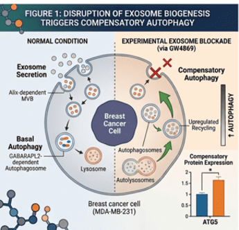
When we blocked the exosome biogenesis pathway in aggressive breast cancer cells (MDA-MB-231), the cells did not simply stop communicating; instead, they upregulated core autophagic machinery, specifically elevating key autophagy-related proteins such as ATG5 and ATG16L1. Conversely, when we pharmacologically inhibited autophagy, the cells compensated by significantly increasing the secretion of exosomes into their surrounding environment. This discovery fundamentally alters our understanding of cancer biology. It proves that multivesicular bodies (MVBs) serve as a central molecular traffic junction, routing cellular cargo toward either lysosomal degradation or extracellular release based on real-time stress signals. Because the cell maintains an active “sensing” system between internal recycling and external secretion, targeting only one side of this equilibrium simply pushes the cell to rely more heavily on the other.
Your research identified the proteins GABARAPL2 and Alix as important regulators. What role do these molecules play, and why could they become promising targets for future cancer therapies?
Our study identified a novel mechanism uniting these two factors. GABARAPL2 acts as a master regulator modulating the levels and secretion of Alix. When autophagy is inhibited, cellular levels of Alix drop while exosome secretion surges; remarkably, knocking down GABARAPL2 prevents this decrease in Alix, highlighting GABARAPL2’s crucial role in governing Alix-dependent exosome packaging and release. Because GABARAPL2 and Alix operate at the intersection of cellular degradation and intercellular signaling, they represent attractive targets for therapeutic intervention. Small molecules or RNA-based therapeutics capable of disrupting the interaction between GABARAPL2 and Alix could shut down the cell’s ability to switch between recycling and secretion, dismantling its primary survival safety net.
Your team found that blocking both pathways together was much more effective than targeting either one alone. Why is this combination strategy so promising?
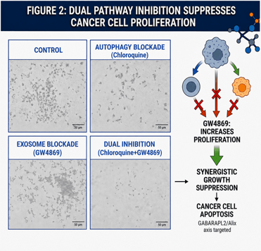
One of the most significant findings of our research is that single-pathway inhibition is largely ineffective at curbing aggressive cancer cell growth. When we blocked exosome biogenesis alone, breast cancer cells actually exhibited an increase in proliferation, driven by compensatory autophagic activity. Similarly, inhibiting autophagy in isolation produced negligible effects on restricting tumor cell growth, as the cells compensated by dumping excess metabolic stress via exosomes.
However, when we implemented a dual-inhibition strategy-simultaneously suppressing both autophagy and exosome biogenesis-the result was a striking reduction in breast cancer cell proliferation.
This combination approach works by trapping the cancer cell in a state of unresolvable metabolic stress. Blocking autophagy prevents the cell from clearing damaged mitochondria and protein aggregates internally, while blocking exosome secretion stops it from expelling waste products or signaling for external support. Unable to clean, recycle, or communicate, the cancer cell experiences metabolic collapse.
How could discoveries like yours contribute to the development of more effective and personalised treatments for breast cancer in the future?
Breast cancer is a highly heterogeneous disease, with subtypes like Triple-Negative Breast Cancer (TNBC) presenting significant clinical challenges due to high recurrence rates and limited targetable receptors. TNBC cells rely heavily on robust autophagy and dense exosome secretion to thrive in nutrient-deprived tumor microenvironments.
Discoveries centering on the GABARAPL2-Alix axis provide a blueprint for precision medicine:
- Biomarker Stratification: Patient tumors can be screened for baseline expression levels of GABARAPL2, Alix, ATG5, and exosomal markers (CD63). High expression profiles could identify patients whose tumors depend heavily on this reciprocal survival loop.
- Combination Drug Cocktails: Instead of administering single-agent autophagy inhibitors (like chloroquine), oncologists could combine autophagy inhibitors with exosome-blocking agents (such as neutral sphingomyelinase inhibitors or novel Alix inhibitors).
- Overcoming Chemoresistance: Standard chemotherapy often induces autophagy as an unintended stress response. By pairing conventional chemotherapy with dual autophagy-exosome blockade, clinicians can prevent cells from developing drug resistance and metastasizing.
Looking ahead, what are the next steps for this research, and what challenges need to be overcome before these findings can eventually benefit patients?
While these findings open exciting possibilities for molecular biology of cancer, several translational hurdles remain before dual-inhibition therapies reach clinical practice. The next immediate step for our research group is validating these cell-culture observations in pre-clinical animal models (in vivo xenografts). We must evaluate whether dual inhibition effectively shrinks tumors and suppresses metastasis in complex physiological environments without causing systemic toxicity. Because healthy cells, particularly neurons, hepatocytes, and immune cells, also rely on basal autophagy and exosome release for physiological maintenance, non-selective systemic inhibition could cause adverse off-target effects. To overcome this challenge, future work will focus on developing targeted drug delivery vehicles, such as antibody-conjugated nanoparticles, designed to deliver dual-pathway inhibitors specifically to tumor tissues. Refining these molecular tools will bring us closer to converting this fundamental biological breakthrough into a viable, life-saving therapeutic reality for cancer patients.