
This article is currently maintained under temporary RFCSR publication support until 13 June 2026.
Imagine you want to catch a local train at a busy Indian railway station during peak hours. The platform is packed, people are everywhere, and boarding the train becomes a real struggle during peak office hours. You need to leave home early, keep extra time in hand, and carefully move through the crowd. Even reaching the train door can take effort because there are simply too many people in a small space. Now compare this with a quiet railway station in the European countryside during a vacation. The station is mostly empty, there is very little crowd, and you can easily walk to your train and board comfortably without stress. Everything moves more smoothly because there is plenty of free space around you.
“Inside living cells, molecular movement is shaped not only by crowding, but also by countless hidden interactions.”
A very similar situation exists when scientists compare the movement of molecules inside a living cell with their movement in a simple test tube experiment. Most biochemical experiments involving proteins, nucleic acids, or small bio-active molecules are usually performed in dilute buffer solutions under standard laboratory conditions. These experiments are extremely important because they provide the first level of understanding about how biomolecules behave. However, these conditions are far from the real environment inside a living cell.
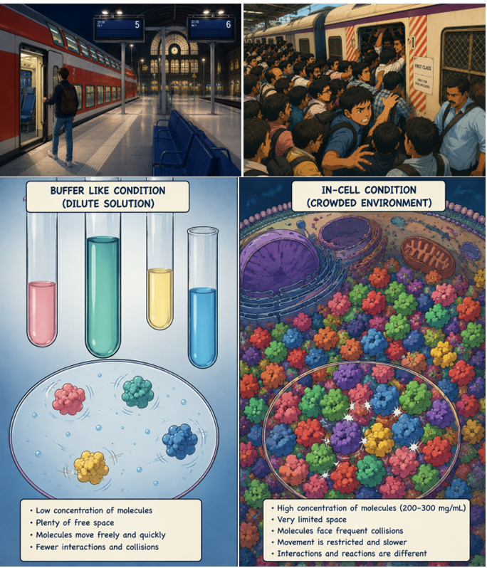
The interior of a cell, especially the cytoplasm, is incredibly crowded —even more severe packed than busiest Indian railway station, one can think of. Inside cells, the concentration of biomolecules can reach nearly 200–300 mg/mL. In such an environment, molecules constantly collide, compete for space, and struggle to move freely. Their behaviour, reaction speed, folding, and interactions can become very different from what we observe in a dilute test tube experiment. Scientists, including me, have tried to mimic this crowded cellular environment in the laboratory by adding artificial “crowder” molecules into buffer solutions, previously. But recreating the true complexity of a living cell is extremely challenging. Even with these efforts, the results often remain incomplete compared with what is observed directly inside living cells. Therefore, experiments performed directly in living cells are critically important for obtaining physiologically relevant insights into molecular behaviour and intracellular transport.
Measuring Diffusion coefficients serves a good proxy to understand the molecular motion in any media including their binding, interactions with the surrounding environments. The classical Stokes-Einstein theory of particle diffusion in ideal solution states that the translational diffusion constant of a particle A (DA) is a function of its dimensions and the solution viscosity.
DA = kBT/6πηrA (1)
where η is the solvent viscosity, kBT is the product of the Boltzmann constant and the absolute temperature, rA is the hydrodynamic radius of the diffusing particle, and DA is a measure of the mean squared displacement per unit time. Interactions with other molecules that lower the mobility will be reflected by lower DA values. Alternatively, if the motion of the molecule is increased due to the presence of another faster moving molecule or the surrounding environment, higher DA values will be recorded. Moreover, inside the complex environment of a cell (presence of lipid microdomains/small organelles may constrain the molecular diffusion), the mean square displacement (MSD) may deviate from linearity with respect to time. This is known as anomalous diffusion. Smaller molecules usually move faster, whereas larger molecules or molecules interacting with surrounding components show slower movement.
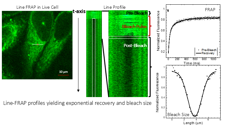
A number of experimental approaches are available to study molecular diffusion both in vitro and inside living cells, although each technique comes with specific advantages and limitations. Fluorescence correlation microscopy (FCS) is considered the gold standard for this purpose; however, its application can be challenging. It requires highly fluorescent molecules (moderate to high quantum yields) and works best at very low concentrations, which may not accurately represent physiological conditions. In addition, immobile or strongly bound molecular populations are often not detected efficiently by this method. Therefore, FRAP is the technique most widely used by experimental biologists. FRAP is relatively simple, minimally invasive, and suitable for studying molecules over a wide concentration range, including biologically relevant conditions. It is particularly useful for molecules that exhibit weak fluorescence signals. Nevertheless, conventional FRAP has limitations, especially when studying rapidly diffusing molecules because of its relatively slow acquisition speed. To address these challenges, I developed an improved high-speed version of FRAP, known as “Line FRAP,” during my postdoctoral research. By using rapid line-scanning instead of conventional imaging modes, the method significantly improves temporal resolution and allows more reliable measurements of fast molecular motion. The use of Line mode greatly improves time resolution of FRAP data acquisition, from 20-100 Hz in the classical mode to 800 Hz in the line mode. This improves data analysis, as intensity and radius of the bleach at the first post-bleach frame is critical. Using this technique, we investigated the diffusion of fluorescently labelled bacterial proteins inside both mammalian and bacterial cells.
Using fluorescently labelled bacterial proteins (molecular weight ~20–100 kDa, bacterial proteins are mostly acidic in nature), we observed that diffusion coefficients in HeLa cells and in E. coli were approximately 2.5-fold and 15-fold lower, respectively, compared with measurements in buffer. This trend is consistent with the progressively increasing degree of cellular crowding from dilute buffer systems to mammalian cytoplasm and finally to the highly compact bacterial cytoplasm, where the available free space for molecular motion becomes substantially reduced. Such crowded intracellular environments influence diffusion through both hard interactions arising from excluded volume effects and soft interactions involving weak, nonspecific molecular associations.
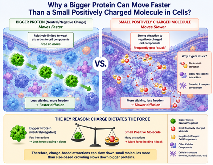
A particularly intriguing finding was that several small molecules (molecular weight <1 kDa) diffused even more slowly than considerably larger proteins within mammalian cells. These results cannot be explained solely by cytoplasmic crowding or occluded volume effects. Although bacterial cytosol is generally more crowded than eukaryotic cytoplasm, mammalian cells possess a much more structurally and chemically heterogeneous intracellular environment due to the presence of multiple membrane-bound organelles, including acidic compartments such as lysosomes.
“Understanding how molecules diffuse inside cells is essential for designing more precise and effective medicines.”
Importantly, weakly basic, amine-containing small molecule drugs (pKa >7) displayed pronounced intracellular “stickiness,” reflected by progressively lower diffusion coefficients with increasing pKa values. This behaviour is consistent with widespread nonspecific electrostatic interactions between positively charged molecules and anionic cellular constituents distributed throughout the cytoplasm. Notably, even after pharmacologically inhibiting lysosomal acidification, the overall diffusion behaviour changed only marginally, suggesting that these nonspecific electrostatic interactions are not restricted to acidic organelles alone but occur broadly across the intracellular environment.
Collectively, these findings indicate that simplified theoretical descriptions such as porous-medium models may adequately describe the diffusion of electronegative larger proteins but are insufficient for explaining the intracellular transport behaviour of positively charged signalling molecules, drugs, peptides, and proteins. For such systems, electrostatic effects and other nonspecific intracellular interactions appear to play a dominant role and therefore need to be incorporated into future models of intracellular diffusion.
Overall, our research is focused on combining advanced optical microscopy and super-resolution imaging approaches to directly visualize small-molecule drugs, their cellular targets, and hidden off-target interactions inside living cells. By uncovering how drugs navigate the complex intracellular environment, we aim to design next-generation precision therapeutic strategies that minimize off-target effects and enable more selective and effective cancer treatments.
Acknowledgements
Dr. Dey acknowledges support from the Department of Biotechnology (DBT), Ministry of Science & Technology, Government of India, through the Ramalingaswami Re-entry Fellowship. ChatGPT (OpenAI) was used for language refinement and generation of selected illustrative graphics. The author reviewed and edited all content and takes full responsibility for the article.


