Tuberculosis is commonly known as a disease affecting the lungs. How can it also affect the brain, particularly in children, and what makes brain tuberculosis a serious medical concern?
Although TB is typically a lung disease, the bacteria can travel through the bloodstream to other organs such as the brain. In the central nervous system, infection can affect the meninges or result in a localized inflammatory mass known as a tuberculoma. A tuberculoma is not cancer, but rather an accumulation of immune cells and damaged tissue that occurs as a result of infection. However, it can be hazardous due to its location. The posterior fossa, which houses the cerebellum and brainstem, is particularly susceptible to the effects of increased volume as even a relatively small amount of increase can block the flow of CSF or compress structures that are critical for movement, consciousness, and breathing. When pressure increases in the skull, the level of consciousness and cognitive conditions of a child can deteriorate quickly. Early recognition is important, not only for infection control, but also to prevent neurological injury from hydrocephalus and mass effect.
Your published case describes a five-year-old child whose brain lesion was initially suspected to be cancer. Could you briefly explain the child’s symptoms, the initial findings, and what made this case particularly unusual?
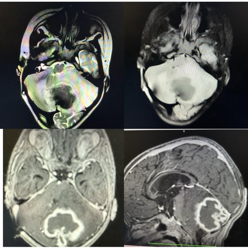
We reported a previously healthy, immunocompetent 5-year-old boy from Kashmir who presented with a progressive increase in headache and irritability over a few weeks. He did not have any known TB contacts, fever, weight loss or night sweats, which would have been suspicious of TB. Brain magnetic resonance imaging showed a large lesion (45 × 38 mm) in the cerebellar vermis and left hemisphere. It compressed the fourth ventricle and surrounding brainstem structures, resulting in obstructive hydrocephalus. Imaging raised concern for a malignant posterior fossa tumor, such as atypical teratoid/rhabdoid tumor (AT/RT) or medulloblastoma. The unique feature was that an intraoperative crush biopsy also indicated an atypical rhabdoid tumor. It was only after the more extensive postoperative tissue examination that it was determined that this seemingly aggressive tumor was actually a tuberculoma.
One of the most striking aspects of your report is that brain tuberculosis closely resembled an aggressive brain tumor. Why can these two conditions look so similar, and what makes it difficult for doctors to distinguish them?
Both a tuberculoma and a malignant tumor can fill space, produce swelling, deform normal anatomy, and obstruct the flow of cerebrospinal fluid. Both can thus cause headache, vomiting, imbalance or other symptoms of increased ICP. They may also have similar MRI appearances. Tuberculomas can appear as solid or ring-like pattern, with surrounding edema and significant mass effect, which are not specific to infection. In our patient, the lesion was mostly hypointense on T2/FLAIR imaging with a hyperintense rim and no diffusion restriction. These observations were useful but did not establish a diagnosis by themselves. Clinical context also makes interpretation difficult: a child can have isolated brain TB without the classic constitutional signs of pulmonary TB. Therefore, imaging should be used to help direct the differential diagnosis, and not as a definitive diagnosis of malignancy or tuberculosis.
In this case, the initial examination of tissue suggested a malignant tumor, but further testing confirmed tuberculosis. What diagnostic methods helped establish the correct diagnosis, and why is detailed laboratory examination so important?
The first crush preparation made intraoperatively was suggestive of a rhabdoid tumor, which is a limitation of rapid tissue assessment: a small, distorted sample may not represent the lesion as a whole. However, multiple permanent histological sections revealed extensive necrosis, epithelioid granulomas, Langhans-type giant cells and macrophages. CD68-positive macrophages, negative epithelial membrane antigen (EMA) staining, and vimentin positivity in fibrotic tissue were demonstrated by immunohistochemistry. Importantly, Ziehl–Neelsen staining revealed acid-fast bacilli, which confirmed the diagnosis of mycobacterial infection in this histological setting. The published report also mentions that the treating institution did not have INI1 staining available when AT/RT was suspected. The lesson is that a preliminary diagnosis should be confirmed by the complete histological picture, appropriate special stains and, if available, molecular assays like Xpert MTB/RIF. This type of testing can avoid sending a child down the wrong path for cancer treatment.
Once tuberculosis was confirmed, the child’s treatment changed significantly. How was the condition managed, and what does the child’s recovery tell us about the importance of early and accurate diagnosis?
The child initially needed a ventriculoperitoneal shunt for obstructive hydrocephalus and subsequently a left suboccipital craniectomy and decompression due to the size of the lesion and its pressure on critical structures. The definitive pathology confirmed tuberculoma and the treatment was changed from the expected oncological pathway to antitubercular therapy. The report shows a nine-month treatment period and a short three-day postoperative course of corticosteroid for edema. Follow-up MRI revealed significant reduction at 3 months and complete radiological resolution at 9 months, and neurological stability during follow-up. This is a positive result, but it does not mean that every child with brain TB should undergo the same surgery or treatment. Treatment is based on the site of the lesions, the presence of hydrocephalus, clinical stability, drug sensitivity and expert opinion. The overall message is that diagnosis is important and affects treatment and prognosis.
Your report highlights that brain tuberculosis may occur even without typical symptoms such as fever, weight loss, or a known history of tuberculosis exposure. What should doctors and parents understand about recognizing this condition, especially in regions where tuberculosis is common?
The lack of fever, weight loss, night sweats, or history of exposure should not rule out CNS tuberculosis, especially in a geographical area where TB is common. Children who have persistent or progressive headache, unexplained irritability, vomiting, gait disturbance, or evidence of increased ICP should be evaluated urgently by a clinician. Tuberculosis should be considered in the differential diagnosis of a posterior fossa mass on imaging, in addition to neoplastic and other infectious diseases. A targeted TB assessment may involve a history of exposure, chest imaging, and immunological tests, but these tests are not sufficient to confirm or exclude a brain tuberculoma. Parents should be aware that these symptoms are not unique to TB or cancer, but are warning signs that need prompt medical attention. The purpose is not to cause anxiety, but to minimize unnecessary delays in the investigation of a potentially treatable condition.
Based on your clinical experience and this case, what are the most important lessons for improving the diagnosis and management of brain tuberculosis in children? What further improvements in medical testing or clinical awareness would help prevent similar diagnostic challenges?
There are three lessons to be learned from our case. First, a wide differential diagnosis should be considered when assessing posterior fossa masses in children, especially in TB-endemic areas. Second, preliminary intraoperative pathology should be interpreted with caution and permanent section histology, with the addition of immunohistochemistry if appropriate, and microbiological stains or molecular tests should be performed. Third, decisions must be made in concert by neurosurgeons, radiologists, pathologists, pediatricians and infectious-disease specialists, as the immediate threat from hydrocephalus may necessitate intervention before the underlying diagnosis is confirmed. Diagnostic uncertainty could be reduced with increased access to rapid molecular testing, appropriate tissue handling and specialist neuropathology. It is also important to teach that isolated CNS TB may be present without pulmonary symptoms. Our report is about one child and does not provide a screening protocol for TB, but does suggest a lower threshold for considering TB and appropriate investigations in children from endemic areas. In the end, diagnostic accuracy saves patients from under- and overtreatment of infection and presumed cancer. It would be helpful to create multidisciplinary review pathways for atypical pediatric brain masses, including explicit reminders to think about infectious causes and to save tissue for microbiological evaluation. Families should be informed that an early radiological or intraoperative impression may be modified following final testing. Follow-up imaging and neurological evaluation are also necessary to document response and complications. The aim is to make a careful and timely diagnosis, while not delaying emergency treatment of raised ICP.