
Looking beyond symptoms
Memory loss is one of the best-known signs of dementia, but it is only the visible part of a much more complex process. Cognitive decline can involve changes in attention, language, planning and everyday function, and different people may reach similar symptoms through different biological pathways. Alzheimer’s disease and vascular dementia are not identical conditions, yet both can be accompanied by disturbances in energy use, amino-acid balance, inflammation and lipid metabolism. Understanding these changes may help researchers develop accessible measures that complement neurological examination, cognitive testing and brain imaging.
“Cognitive decline may be accompanied by interconnected changes in energy metabolism, amino-acid balance, inflammation and lipid-associated signals.”
Why metabolism matters to the brain
The brain has a high and continuous demand for energy. Neurons depend on carefully regulated supplies of mainly glucose and other fuels, functioning mitochondria, and a balanced environment of amino acids and lipids. Inflammation and vascular or metabolic comorbidities can influence these systems. Metabolomics—the systematic study of small molecules in biological samples—offers a way to examine the combined biochemical consequences of genes, lifestyle, ageing and disease. Blood is particularly attractive because collection is relatively straightforward and can be repeated over time. However, a blood profile reflects the whole body; it should not be treated as a direct readout of brain chemistry.
From mild cognitive impairment to dementia: evidence from NMR
Our work using proton nuclear magnetic resonance (¹H NMR) spectroscopy has examined circulating metabolic features across different stages of cognitive impairment. In a 2021 study of a North Indian population, plasma from a subset of participants with mild cognitive impairment and healthy controls was profiled using high-field ¹H NMR. Lysine, glycine and glutamine emerged as a candidate three-metabolite panel. The study also considered diffusion tensor imaging and cognitive assessment, placing metabolic findings alongside clinical and brain-imaging information. These results supported further investigation of blood-based metabolic markers, but the sample analyzed by NMR was modest and the findings require validation in independent populations.
A broader metabolic signature in cognitive decline
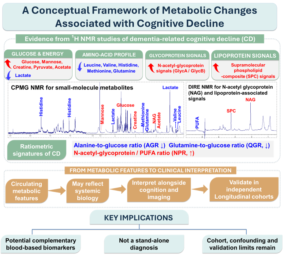
In our more recent study published in Biophysical Chemistry, serum samples from 40 participants with cognitive decline and 54 cognitively normal controls were analysed using 800 MHz ¹H NMR spectroscopy. Thirty-three metabolites and selected metabolic ratios were quantified. The cognitive-decline group showed higher glucose, pyruvate, acetate, creatine and mannose, alongside lower levels of several features including lactate, leucine, valine and histidine. The glutamine-to-glucose ratio (QGR) and alanine-to-glucose ratio (AGR) were also lower. AGR was positively associated with Mini-Mental State Examination scores, while AGR and QGR declined with increasing severity in the study cohort. QGR, AGR and the histidine-to-tyrosine ratio showed promising discrimination within that cohort.
Why look at glycoproteins and lipoproteins as well?
Small metabolites are only one part of the circulating biochemical picture. In a separate study currently under review at Clinica Chimica Acta, our team has explored complementary NMR experiments designed to capture both low-molecular-weight metabolites and broader glycoprotein- and lipoprotein-associated signals. The submitted analysis reports higher N-acetyl-glycoprotein signals and supramolecular phospholipid-composite signals in the cognitive-decline group, together with changes in glucose, mannose, amino acids and metabolic ratios. These findings are preliminary until peer review and independent validation are complete. Nonetheless, they illustrate the value of asking whether several biochemical layers—rather than a single metabolite—can provide a more informative picture of systemic changes accompanying cognitive decline.
What these findings mean—and do not mean
Taken together, the studies support a view of cognitive decline as a condition that may be accompanied by interconnected metabolic changes, rather than a process understood only through memory symptoms. Ratios such as QGR and AGR may be useful because they express relationships between metabolites, not simply the level of one molecule. Glycoprotein-associated signals may add information about systemic inflammatory activity, while lipoprotein-related signals may reflect changes in lipid transport and composition. These are biological interpretations, not proof that a specific pathway caused cognitive impairment. Cross-sectional associations cannot establish whether metabolic changes precede cognitive decline, result from it, or arise partly from other conditions.
The challenges before clinical use
Several hurdles must be cleared before these observations can become clinical tests. First, findings need replication in larger, independent and diverse cohorts, including participants with different educational backgrounds, diets, vascular risks and comorbidities. Second, longitudinal studies should determine whether metabolic profiles change as cognition changes and whether they can predict conversion from mild cognitive impairment to dementia. Third, studies need careful control of factors such as age, sex, fasting status, medication, diabetes, kidney and liver function, sample handling and storage. Dementia is heterogeneous, and a marker that performs well in one group may not generalize to another.
Analytical and statistical standards matter equally. NMR offers reproducible profiling and can measure multiple metabolites in one experiment, but some spectral signals overlap and assignments may remain putative unless confirmed with additional methods. Multivariate models can appear more accurate than they truly are if data are over-fitted or validation is incomplete. Candidate markers should therefore be evaluated with transparent preprocessing, correction for multiple testing, suitable internal validation and external testing. Ultimately, their value must be compared with established clinical assessments and available blood, cerebrospinal-fluid or imaging biomarkers—not merely with healthy controls.
A practical path forward
The next step is not to promote a metabolic panel as a stand-alone diagnostic test, but to test whether it adds useful information to existing care. Prospective multicenter studies could combine standardized clinical phenotyping, cognitive scores, brain imaging and established biomarkers with serum or plasma NMR profiles. Where feasible, metabolomics could be integrated with lipidomics, proteomics and other data layers, using machine-learning models that are independently validated and interpretable. Researchers should also examine whether metabolic patterns differ by dementia subtype and whether they reflect modifiable contributors such as vascular risk or nutritional status. The conceptual framework is presented in Figure1.
Key Takeaways
A diagnosis of dementia must remain grounded in a careful clinical evaluation. A blood test based on metabolomics is not currently a substitute for that process. Yet the ability to measure many metabolic features from a small sample offers a promising research route to understanding the biology that accompanies cognitive decline. Our studies—from mild cognitive impairment to broader serum metabolite profiles and exploratory glycoprotein/lipoprotein signals—add to this developing evidence base. The most important task now is rigorous validation and collaboration between neurologists, analytical scientists, statisticians and clinical researchers. Looking beyond memory loss does not mean looking away from patients; it means seeking a fuller biological understanding that may, in time, support more precise assessment and monitoring.