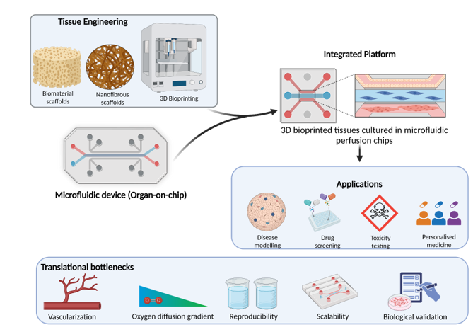
Biomedical engineering is increasingly at the interface of living and engineered systems. Many biological questions are still difficult to answer using standard two-dimensional (2D) cell culture. Cells in 2D cultures lack the three-dimensional (3D) architecture, cell-cell interactions, extracellular matrix cues, and dynamic physical environment that would be present in a tissue in the body. Animal models are useful, but there are inevitably differences between species that can limit the translation of findings from the animal to the human. This has led to the development of experimental systems that can mimic some of the features of human tissues, while being measurable, controllable, and amenable to engineering design. Tissue engineering and organ-on-a-chip technologies are two approaches to this, which take a very different perspective. Tissue engineering involves the combination of cells, biomaterials, and biochemical or physical cues to generate a tissue-like environment. Organ-on-a-chip systems use micro-fabricated devices and precisely controlled micro-environments to recreate aspects of organ physiology such as fluid flow, mechanical stimulation, and cell-cell interactions. The two approaches together provide a much wider range of opportunities to study disease, assess potential therapies, and explore regenerative strategies.
“Tissue engineering and organ-on-a-chip technologies are complementary: one organizes cells and materials, while the other provides controlled environments to investigate tissue function”
Engineering the cellular microenvironment
A tissue is more than just a aggregation of cells. Its behavior is determined by the ECM (extracellular matrix), cellular architecture, biochemical signals, and mechanical properties of the tissue. Biomaterials and scaffolds provide a framework for cells to attach to, communicate with each other, and perform their tissue-specific functions. The materials used must therefore be selected with regard to the specific biological application in mind. The research from my lab is a good example of this. We examined soy protein isolate silk-fibroin nanofibrous scaffolds and evaluated their potential for skin tissue regeneration. This involved the characterization of the scaffolds and assessment of their wound healing potential in an animal model (Varshney et al., 2020). We have also examined psyllium husk-gelatin blends as potential bioinks and used rheological and mechanical characterization to assess those (Biofabrication and 3D bioprinting take this design process further by enabling the precise spatial positioning of cells and biomaterials. However, printability is not the same as biological suitability. A formulation must be printable but also must maintain cell viability, allow for the transport of oxygen and nutrients, and allow for tissue maturation. Rheological and mechanical characterization in conjunction with biological testing is therefore a necessary step. Our FRESH (Freeform Reversible Embedding of Suspended Hydrogels) work demonstrated the use of a chitosan bio-ink to produce complex geometries, including a human tri-leaflet heart valve and a section of a coronary arterial tree . The use of a temporary embedding medium to support the printed structure allows for the maintenance of complex geometries during the fabrication process. This work illustrates how engineering approaches can be used to expand the range of geometries possible with hydrogel-based materials.
Organ-on-a-Chip technologies
Organ-on-a-chip systems combine micro-engineering with cell culture via the use of micro-channels and chambers to control fluid flow, create interfaces between different cell populations, and deliver precisely defined chemical and mechanical stimuli. The degree to which a given system can mimic the relevant features of a particular organ (e.g., liver, lung, intestine, and nervous tissue) will vary according to the design of the system. The importance of being able to control the physical conditions is nicely illustrated by our earlier work using a microfluidic system to study surfactant secretion in alveolar type II epithelial cells. In the study, we revealed that the level of flow-induced wall shear stress could be used to modulate aspects of the surfactant secretory process in these cells, with the effect being different in different cell models. That study is a good example of one of the key benefits of the microfluidic approach, which is, the ability to systematically vary a physical stimulus and observe the resulting cellular response in a controlled manner. Organ-on-a-chip systems are increasingly used for drug screening, toxicity testing, and disease modelling. The effectiveness of the cells depends on their quality, the physiological functions they can reproduce and the validation of these functions against relevant biological references. In this context, studies of brain-on-a-chip demonstrate how microfluidic engineering can advance neuronal modelling, in collaboration with my group, developed a microfluidic platform enables the incorporation of 3D neuronal networks for site-specific neuro-pharmacological treatment and optical monitoring of neuronal activity. Another study by our Poddar et al. (2019) and my group reported on compartmentalized microfluidic devices for low-density primary neuronal culture. This study provided the possibility to study neuronal growth, axonal development and cellular interactions. Together, these studies highlight the potential of microfluidic technologies for creating more functionally relevant and tightly controlled neuronal models for biomedical research and drug discovery.
Organ-on-a-chip systems are being used for a range of applications, including drug screening, toxicity testing, and disease modelling. Their usefulness in any of these areas will depend on the quality of the cells used, the physiological functions that are being represented, and the extent to which they have been validated against appropriate biological references. It is important to note that a chip is not a miniaturized version of a whole human organ. Most systems can only replicate a subset of the features of the organ in question, and other functions may be greatly simplified or entirely absent. The application of the technology and the degree to which it is reproducible will be paramount.
Where the technologies meet
The combination of tissue engineering and organ-on-a-chip design is a promising avenue. Engineered scaffolds and bioprinted tissues can provide the spatial organization and matrix cues that are important, and microfluidic platforms can provide the controlled flow, dynamic stimulation, and defined interfaces between cell populations. The potential is that you can then go on to study how the engineered tissue responds to a more physiologically relevant environment. Our own work in the area of liver tissue engineering is a case in point. We recently looked at the performance of lyophilized, electrospun, and 3D-printed psyllium husk polysaccharide-based scaffolds in the culture of primary rat hepatocytes. All the scaffolds supported cell viability, aggregate formation, and the secretion of liver-specific functional markers (albumin and urea). The level of functional performance was dependent on the scaffold fabrication method. This highlights the need to go beyond simply looking at the look and feel of the tissue and the physical structure to also assess relevant biological function. When combined with microfluidic control, such scaffolds could form the basis for studying the impact of matrix architecture, fluid flow, and cell organization on tissue function. More generally, engineered liver and neural tissues could eventually be used to study selected organ-organ interactions in both health and disease. The added value of such integrated platforms will of course have to be demonstrated through direct experimentation and comparison to appropriate reference models rather than simply being assumed from their complexity.
For biomedical engineering, this convergence means that we need to bring together materials science, cell biology, microfabrication, fluid mechanics, imaging, and quantitative analysis around a common biological question. You have to define what function the model is going to be trying to reproduce, you have to define how you’re going to measure that function, and you have to have the evidence that it’s actually doing it.
Translational bottlenecks
There are still a whole host of issues in this area. With thick tissues, you can easily get gradients of oxygen and nutrients unless you have appropriate transport mechanisms in place In thick engineered tissues, oxygen and nutrient gradients can readily develop unless adequate transport mechanisms are incorporated to ensure sufficient distribution throughout the tissue. . Vascularization, long-term maturation, and maintenance of tissue-specific function are all still issues. There is a lot of variability in the cell sources, biomaterial batches, fabrication conditions, and device geometries, which will all have an impact on reproducibility. For comparison of results between labs to be meaningful, it is absolutely essential that there are standard protocols and shared benchmarks. There is a need to strike a balance between model complexity and cost, throughput, ease of use, and scalability. You cannot simply assume that more complex models are necessarily more biologically relevant. You need to validate the model for the application of interest. Progress in this area will require working closely with clinicians, regulatory scientists, and end users to ensure that the platforms we develop are actually going to address some real biomedical problems.
Future prospects for more predictive bioengineering
Tissue engineering and organ-on-a-chip technologies are revolutionizing biomedical research by allowing the creation and study of human-relevant biological systems in a more accurate and precise manner. The aim is not simply to build tissue-like structures or miniaturized organs, but to develop tools that can answer biological questions and, when properly validated, inform decisions about therapy and regenerative medicine. In the field of biomedical engineering, the opportunity is to combine engineering control with biological insight and to judge the success of an innovation on the basis of the quality, reproducibility, and relevance of the knowledge that it generates.
A call to action: From engineering innovation to biomedical impact
To truly unlock the potential of tissue engineering and organ-on-a-chip, we need to go beyond individual technologies and work towards integrated, reproducible, and biologically validated systems. The next step involves strengthening collaboration among bioengineers, cell biologists, clinicians, and regulatory scientists to establish shared standards for model validation and ensure that emerging platforms are scalable, reliable and suitable for biomedical applications.