When life-saving medicines stop working
Imagine having an infection that was once easily treated with antibiotics, but the same medicines no longer work. This is one of the growing challenges facing modern medicine. Antibiotic resistance occurs when bacteria develop the ability to survive medicines designed to kill them or stop their growth.Antibiotics have transformed healthcare, making previously dangerous infections treatable and enabling safer surgeries, cancer treatments and organ transplantation. However, their effectiveness is increasingly threatened by antimicrobial resistance (AMR), which makes infections harder to treat and increases the risk of serious illness.
But how can a microscopic bacterium survive a medicine designed to destroy it? The answer lies partly in the remarkable adaptability of bacteria. Some prevent antibiotics from entering their cells, others pump medicines out, while some produce specialised proteins called enzymes that destroy or inactivate antibiotics. Understanding these enzymes is an important step towards understanding antibiotic resistance.
Sometimes, the key lies in something invisible to the naked eye: the three-dimensional structure of a protein.
“A larger active site does not necessarily make an enzyme more effective; subtle molecular changes can reshape how it interacts with antibiotics.”
Bacterial enzymes: Tiny machines with a powerful role
Enzymes are proteins that accelerate chemical reactions in living organisms. They perform essential tasks, from helping us digest food to enabling bacteria to grow and survive. However, some bacterial enzymes work against antibiotics. A particularly important group is called beta-lactamases.
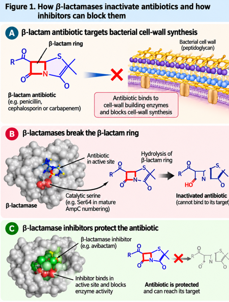
Penicillins, cephalosporins and carbapenems belong to a major family of antibiotics known as beta-lactams. These medicines work by interfering with the construction of the bacterial cell wall, an essential protective structure. Beta-lactamases defeat this strategy by breaking open a critical chemical ring in these antibiotics, rendering them inactive (Figure 1A).
Think of an antibiotic as a key designed to interrupt an essential process in a bacterium. A beta-lactamase acts like a tool that damages that key before it can do its job.
Beta-lactamases are diverse and are commonly grouped into four classes (A, B, C and D) based on their molecular characteristics. Most use an amino acid called Serine64 to break down antibiotics, whereas class B enzymes use zinc ions to facilitate the reaction. Understanding these different mechanisms has been central to developing strategies to counter antibiotic resistance (Figure 1B).
The development of beta-lactamase inhibitors represents an important advance in this field. Earlier inhibitors, such as clavulanic acid, sulbactam and tazobactam, helped restore the activity of certain beta-lactam antibiotics against bacteria producing susceptible enzymes. Newer inhibitors, including avibactam, relebactam and vaborbactam, have expanded the range of beta-lactamases that can be targeted (Figure 1C). However, no single inhibitor is effective against every enzyme, and the continuing emergence of new variants presents an ongoing challenge.
One important example is Pseudomonas aeruginosa, a bacterium that can cause serious infections, particularly in vulnerable patients. It produces enzymes called Pseudomonas-derived cephalosporinases (PDCs), which belong to the AmpC family of beta-lactamases. As these enzymes evolve, their interactions with different antibiotics may also change. Understanding these changes requires more than knowing which genes a bacterium carries; we need to understand the proteins those genes produce.
Why does the shape of a protein matter?
Proteins are not rigid objects. They fold into intricate three-dimensional structures, and their shapes help determine what they can do. Many enzymes contain an active site, a specialized region where a chemical reaction takes place. In beta-lactamases, this region helps recognize and process susceptible antibiotics.
Imagine a workspace designed for a particular task. Its size, shape, chemical properties and flexibility all influence how efficiently the task can be completed. An enzyme’s active site works in a similar way.
Changes in the amino acids that make up a protein can alter the shape or chemical environment of its active site. These changes may influence how an antibiotic enters, how it is positioned and how efficiently the enzyme breaks it down. Importantly, a larger active site does not automatically mean a more effective enzyme, just as a smaller workspace does not necessarily make every task impossible. The outcome depends on the particular antibiotic and the precise arrangement of the enzyme’s components.
This is where structural biology becomes valuable. Techniques such as X-ray crystallography allow researchers to determine the three-dimensional structures of proteins and investigate how molecular changes may influence their function. When combined with biochemical experiments and computational approaches, structural information provides a more complete picture of how enzymes interact with antibiotics.
A closer look at PDC-16: What our research revealed
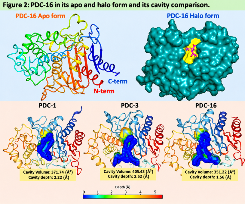
Our recent research focused on PDC-16, a variant of the AmpC beta-lactamase associated with carbapenem-resistant P. aeruginosa. Published in the International Journal of Biological Macromolecules (2026Sep 30;383), our study examined the enzyme’s structure and function to understand how its molecular architecture relates to its activity against different antibiotics. Using X-ray crystallography, we determined two structures: PDC-16 without an antibiotic bound and PDC-16 with imipenem, a carbapenem antibiotic, bound within its active site (Figure 2). X-ray crystallography uses the diffraction of X-rays by protein crystals to reveal the arrangement of atoms in a protein. In simple terms, it allows scientists to build a detailed three-dimensional picture of a molecular machine that is far too small to see directly.
The findings revealed an interesting feature of PDC-16. Its active-site cavity was more constrained than those of the related enzymes used for comparison. Our structural analysis estimated its cavity volume at approximately 351 Å3, compared with 372 Å3 for wild-type PDC-1 and 405 Å3 for cavity expanded PDC-3. These measurements describe the space available within the enzyme’s active site (Figure 2).
This observation is interesting because structural adaptation in beta-lactamases does not always involve an expansion of the active site. Changes in the positions of amino acids and the flexibility of nearby protein regions may also influence how an enzyme interacts with different antibiotics. The relationship between active-site architecture and enzyme function is therefore more complex than simply having a larger or smaller cavity.
We also observed structural rearrangements in PDC-16 when imipenem was bound. The structure captured a snapshot of the enzyme–antibiotic interaction, including a covalent acyl-enzyme intermediate. Although this provides valuable information about the interaction, further studies are needed to understand the precise molecular movements that occur during the catalytic process.
Our biochemical experiments added another important dimension. PDC-16 displayed substrate-dependent kinetic behaviour, with differences in its apparent affinity and catalytic activity across the beta-lactam antibiotics examined. Its behaviour towards one antibiotic could not simply be used to predict its behaviour towards another.
This is an important lesson: antibiotic resistance cannot always be explained by labelling an enzyme as simply powerful or weak. Its effects depend on the antibiotic, the molecular features of the enzyme and the broader biology of the bacterium. These findings do not mean that PDC-16 alone explains carbapenem resistance. Other mechanisms, including reduced antibiotic entry and the activity of drug-efflux pumps, can also contribute.
From understanding enzymes to finding better treatments
If enzymes can help bacteria defeat antibiotics, could we develop medicines that stop these enzymes from working?
One strategy is to identify beta-lactamase inhibitors. These compounds interfere with the enzyme, allowing a partner antibiotic to retain its activity against bacteria that would otherwise be able to inactivate it. However, different beta-lactamases respond differently to inhibitors, making it important to understand the molecular features that determine inhibitor recognition.
Our study explored this challenge using computational approaches to identify compounds that might interact with the PDC-16 active site. Although some candidates showed promising predicted interactions, subsequent biochemical experiments demonstrated that favourable computational binding predictions did not necessarily translate into effective enzyme inhibition.
This distinction is crucial. A molecule may appear to fit into an enzyme’s active site on a computer but still fail to block its activity in the laboratory. Computational screening is therefore a useful starting point, not proof of inhibitor activity. Candidate molecules must undergo experimental testing to establish whether they inhibit the enzyme, can reach their target inside bacterial cells, are safe and offer meaningful benefits in infection models and, ultimately, clinical studies.
Future perspectives: Where can structural biology take us?
The future of antibiotic resistance research depends on connecting molecular discoveries with practical medical needs.
First, studying more beta-lactamase variants will help researchers understand how bacterial enzymes adapt to different antibiotics. Comparing their structures, activities and evolutionary changes may reveal patterns that are difficult to identify from genetic information alone.
Second, advances in structural biology, computational modelling and artificial intelligence could help scientists identify promising inhibitor candidates more efficiently. These approaches may guide the design of molecules that interact with important regions of an enzyme, including regions that differ between variants. Third, researchers must investigate how these enzymes function inside living bacteria. A purified protein studied in the laboratory provides valuable information, but bacterial cells are complex systems. Membrane permeability, efflux pumps, gene regulation and other resistance mechanisms can influence whether a treatment succeeds.
Finally, collaboration among structural biologists, microbiologists, medicinal chemists, clinicians and public-health researchers will be essential to translate molecular discoveries into useful treatments.
Conclusion: Small molecular changes, big consequences
Antibiotic resistance is a global challenge, but understanding it begins with fundamental scientific questions: How does a bacterial enzyme recognise an antibiotic? How do small changes in its structure influence its activity? And can these insights help us find better ways to block it? Our research on PDC-16 illustrates why these questions matter. Its constrained active-site architecture and substrate-dependent kinetic behaviour highlight the complexity of the relationship between protein structure and antibiotic resistance. Structural biology does not immediately produce new medicines, but it provides essential molecular knowledge that can guide future research and the search for better therapeutic strategies.