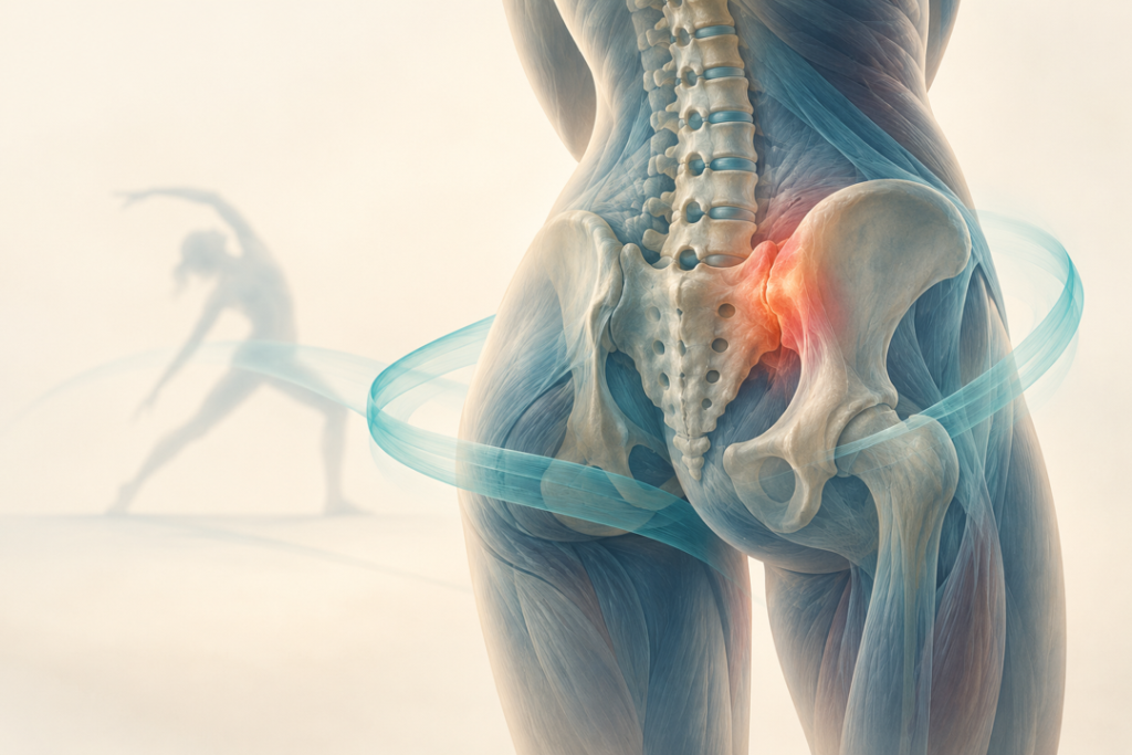
Sacroiliac (SI) joint pain is considered a significant contributor to chronic low back pain, accounting for nearly one-quarter of cases among adults with prominent symptoms. Unlike typical midline low back pain, discomfort arising from the SI joint is generally felt on one side and occasionally affects both sides. Women appear to be affected more frequently than men, which may be partly related to differences in pelvis biomechanics. The SI joint in women allows greater mobility, potentially increasing mechanical stress, loading demands, and strain on the supporting pelvic ligaments. These biomechanical reasons may help explain why SI joint dysfunction is more common during pregnancy and the postpartum period, when substantial changes occur in pelvic structure and loading.
Pain associated with SI joint dysfunction is often experienced not only in the lower back but may also extend downward toward the leg. Functional movements that can intensify these symptoms, include transitions from seated to standing or from lying down to standing. Activities that place greater mechanical demand on the pelvis, such as climbing up or walking down stairs, may also aggravate the pain.
When the Pain Doesn’t Tell the Whole Story
SI joint pain can be a real diagnostic puzzle. The joint sits deep within the pelvis, and pain arising from this region can appear in different ways and may overlap with symptoms from the lower back, hip, or surrounding muscles. This makes it difficult to identify the SI joint as the true source of pain based on the symptoms alone.
So, how do clinicians solve the puzzle?
A careful clinical assessment is the first step. The physiotherapist looks at the patient’s history, movement patterns, and areas of tenderness around the SI joint. Although commonly used assessments, such as walking pattern observation and the Trendelenburg test, can provide useful information, they may also be influenced by problems outside the SI joint and, therefore, cannot be relied upon alone.
This is where the provocation test becomes particularly valuable. Tests such as Gaenslen’s thigh thrust, distraction compression, and FABER test are designed to place stress on the SI joint and determine whether the patient’s familiar pain can be reproduced. Rather than depending on one test, clinicians often consider the findings from several tests together to build a clearer clinical picture.
When the diagnosis remains uncertain, image-guided injection of a local anesthetic into the SI joint has traditionally been used as a reference method to determine whether the joint contributes to pain.
The key message is that a single test cannot always identify SI joint pain. However, a combination of patient history, physical examination, pain-provocation tests, and appropriate investigations can help clinicians move from uncertainty towards a more confident and individualized rehabilitation plan.
Treating SI Joint Pain: Back to Basics
Once SI joint dysfunction is identified, physiotherapy plays an important role in restoring movement and reducing pain. Traditionally, treatment has focused on a combination of therapeutic exercise, manual therapy, joint mobilisation, stretching, strengthening, massage, patient education, and physical modalities such as heat, ultrasound and TENS.
The aim is not simply to reduce pain, but to improve mobility, stability, strength, and confidence in everyday movement. However, SI joint related symptoms are often grouped under the broad label of low back pain, which can make specific management challenging. This is why a more focused physiotherapy approach is gaining attention, recognizing the SI joint as a potential source of symptoms and tailoring treatment to the individual rather than treating every case as routine back pain.
Looking beyond Traditional Treatment
For young physiotherapists and researchers stepping into the field of SI joint pain, the future belongs to those willing to look beyond traditional boundaries. Rehabilitation is gradually moving towards a more integrated approach that combines clinical reasoning, biomechanics, exercise therapy, manual therapy, and emerging therapeutic technologies. Approaches such as laser therapy, induction therapy, matrix-rhythm therapy, magnetotherapy, shockwave therapy, manual therapy, and myofascial therapy using tools, manual, as well as self-administered techniques are attracting growing interest as potential components of comprehensive rehabilitation. However, innovation must always be guided by scientific evidence and patient needs. SI joint pain can be complex, and no single treatment approach is likely to work equally well for every patient.
The next generation of physiotherapists should therefore focus on evidence-based practice, not only on learning new techniques but also on understanding when, why, and for whom a particular intervention should be used. Careful assessment, clinical reasoning, patient education, individualized exercise, and continuous evaluation should remain at the heart of rehabilitation.
The future will also depend on collaboration. When clinicians, physiotherapists, researchers, and technology developers work together, new possibilities can emerge for improving pain management, mobility, function, and quality of life. For young researchers, this is an exciting time to contribute to a field that is constantly evolving.
Ultimately, the goal is not simply to introduce newer modalities, but to make rehabilitation safer, more effective, evidence-based, and truly patient-centered. The coming years will bring important advances in how SI joint pain is assessed and managed, and young physiotherapists and researchers will play a key role in shaping that future.