Skeletal muscle is one of the largest organs in the human body and plays a critical role not only in movement and posture but also in maintaining metabolic health. It is the primary site of insulin-stimulated glucose disposal, accounting for more than 75% of insulin-mediated glucose uptake. Consequently, skeletal muscle is a major regulator of whole-body glucose homeostasis and is among the first tissues to develop insulin resistance. These functions underscore its central importance in maintaining metabolic balance and overall health.
The functional units of skeletal muscle are multinucleated cells known as muscle fibers or myofibers. Skeletal muscles contain different types of myofibers, each specialized for distinct physiological functions. Broadly, they are classified into slow-twitch (Type I) and fast-twitch (Type II) fibers.
Slow-twitch fibers contract relatively slowly but are highly resistant to fatigue. Rich in mitochondria, they generate energy primarily through oxidative phosphorylation, making them ideally suited for endurance activities such as walking, cycling, and long-distance running (Figure 1). In contrast, fast-twitch fibers contract rapidly and generate greater force but fatigue more quickly. They rely predominantly on glycolysis to meet their energy demands and are essential for explosive activities such as sprinting, jumping, and lifting heavy loads (Figure 1). Fast-twitch fibers are further classified into subtypes that differ in their contractile and metabolic properties.
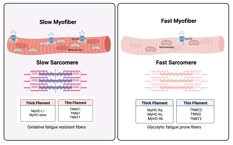
Muscle fiber identity is determined by the specific myosin heavy chain (MyHC) proteins they express. Slow fibers express MyHC-slow, whereas fast fibers express MyHC-IIa, MyHC-IIx, or MyHC-IIb (Figure 1). The relative abundance of these fiber types varies among individuals and is influenced by genetics, age, physical activity, and training. Importantly, skeletal muscle remains highly adaptable throughout life, with regular exercise modifying both the metabolic and functional characteristics of individual fiber types.
Among all muscle fiber types, slow myofibers exhibit the greatest capacity for glucose uptake because they express higher levels of the glucose transporter GLUT4 (Figure 1). Recent studies have shown that exercises specifically targeting slow muscles, such as the soleus push-up, can significantly improve whole-body glucose regulation. These observations highlight the untapped therapeutic potential of skeletal muscle and particularly slow-twitch fibers in preventing and treating metabolic diseases.
“Skeletal muscle is not merely a tissue for movement—it is one of the body’s most important regulators of blood sugar and metabolic health”
Why Your Muscles Matter for Managing Blood Sugar?
Diabetes has emerged as one of the fastest-growing global health challenges, with India bearing one of the world’s largest disease burdens. Physical inactivity and prolonged sedentary behaviour are major contributors to insulin resistance and type 2 diabetes. Yet the role of skeletal muscle in regulating metabolism often receives far less attention than organs such as the pancreas or liver.
Extended periods of inactivity impair the metabolic function of skeletal muscle by reducing glucose uptake, disrupting insulin signalling, and compromising mitochondrial function. Because skeletal muscle is responsible for clearing most of the glucose from the bloodstream after a meal, even modest declines in muscle health can substantially affect blood glucose regulation. These observations emphasize that maintaining healthy skeletal muscle is fundamental to preventing metabolic disease.
Discovering a New Link Between Muscle Health and Diabetes
A recent study from our laboratory has uncovered a previously unknown molecular mechanism linking skeletal muscle health to whole-body metabolism. The work, recently published in Science Advances, demonstrates how the loss of a single muscle-specific contractile protein can directly trigger insulin resistance and metabolic dysfunction.
Our study identifies a previously unrecognized role for MyHC-slow, the contractile protein encoded by the Myh7 gene, in maintaining systemic metabolic health. Beyond its well-established function in muscle contraction, MyHC-slow appears to regulate mitochondrial function, antioxidant defence, and glucose metabolism through the NRF2 signalling pathway, revealing an entirely new connection between muscle structure and metabolic regulation.
Individuals with type 2 diabetes often exhibit a reduction in slow muscle fibers and decreased expression of MyHC-slow. To determine whether this loss contributes directly to disease development, we generated genetically engineered mice lacking MyHC-slow specifically in skeletal muscle. This model enabled us to investigate the biological consequences of losing this single contractile protein.
What Happens When MyHC-slow Is Lost?
Eliminating MyHC-slow from skeletal muscle initiated a cascade of pathological changes that closely resembled the early features of type 2 diabetes.
Skeletal muscle atrophy: Loss of MyHC-slow resulted in selective degeneration of slow-twitch fibers, muscle atrophy, reduced muscle mass, diminished force production, and impaired muscle function.
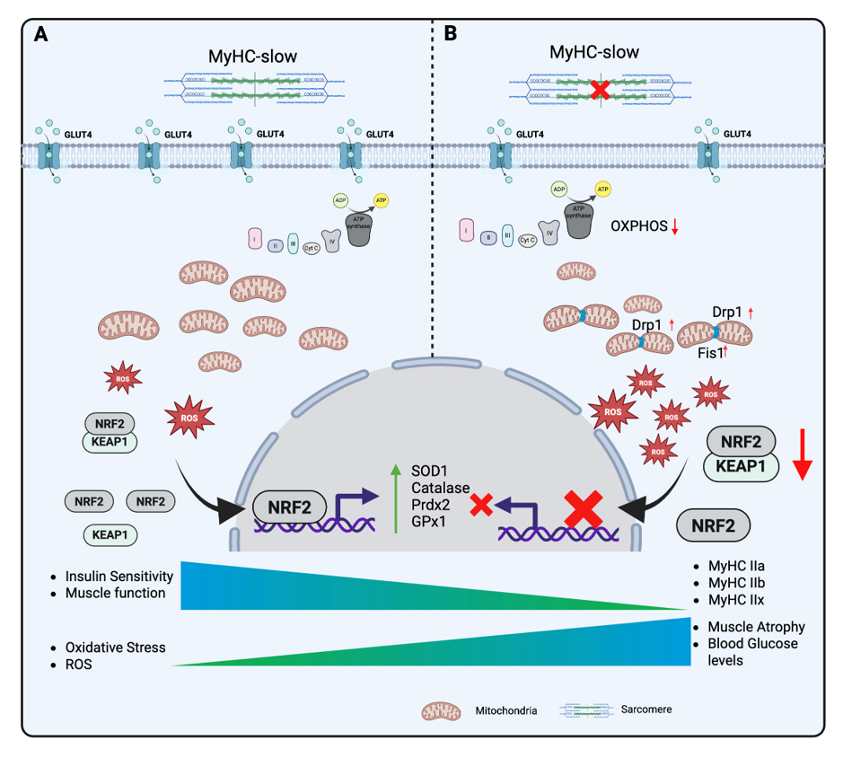
Mitochondrial dysfunction and oxidative stress: The absence of MyHC-slow severely disrupted mitochondrial structure and function, leading to excessive production of reactive oxygen species (ROS). The resulting oxidative stress compromised cellular energy production and impaired muscle metabolism (Figure 2).
Reduced glucose uptake and insulin resistance: Diseased muscle showed a marked reduction in GLUT4 expression, limiting insulin-stimulated glucose uptake. This defect ultimately led to systemic insulin resistance and impaired blood glucose control (Figure 2).
Suppression of the NRF2 antioxidant pathway: One of the most striking findings was the profound reduction in activity of the NRF2 signalling pathway, the master regulator of cellular antioxidant defence. Reduced NRF2 activity weakened the muscle’s ability to neutralize ROS, further amplifying oxidative damage, mitochondrial dysfunction, and metabolic failure (Figure 2).
Therapeutic Targeting: Restoring the NRF2 Antioxidant Pathway
Having identified suppression of NRF2 as a key event, we investigated whether restoring this pathway could rescue muscle function. We treated the MyHC-slow-deficient mice with sulforaphane, a naturally occurring compound abundant in cruciferous vegetables such as broccoli that is known to activate NRF2.
The results were highly encouraging. Activation of NRF2 reduced oxidative stress, restored mitochondrial function, improved muscle health, and significantly rescued the metabolic defects caused by the loss of MyHC-slow. These findings identify the NRF2 pathway as a promising therapeutic target for metabolic disorders characterized by skeletal muscle dysfunction.
The Bigger Picture
For many years, muscle contraction and metabolism were viewed as largely independent biological processes. Our study challenges this traditional view by demonstrating that the proteins responsible for muscle contraction also play fundamental roles in regulating cellular metabolism and whole-body glucose homeostasis.
These findings reveal that skeletal muscle is not merely a tissue for movement but also a critical regulator of metabolic health. By uncovering a molecular link between muscle structure, mitochondrial function, antioxidant defence, and insulin sensitivity, our work opens new opportunities for developing therapies for diabetes, metabolic syndrome, and muscle-wasting disorders.
Looking Ahead
Our findings raise several important questions that will guide future research.
First, it will be essential to determine whether similar mechanisms operate in humans. Analysing skeletal muscle biopsies from individuals with type 2 diabetes, metabolic syndrome, and age-related muscle loss (sarcopenia) will help establish the clinical relevance of MyHC-slow and NRF2 in metabolic disease.
Second, although sulforaphane showed considerable promise in our preclinical studies, further work is needed to evaluate its long-term safety, pharmacological properties, and clinical efficacy. The development of next-generation NRF2 activators with greater muscle specificity could provide even more effective therapeutic options.
Finally, an important unanswered question is how the loss of a structural muscle protein leads to suppression of NRF2 signalling. Understanding the molecular communication between the sarcomere, mitochondria, and the nucleus including mechanotransduction pathways and epigenetic regulation will provide deeper insight into how skeletal muscle controls whole-body metabolism. Together, these studies have the potential to reshape our understanding of skeletal muscle biology and establish new strategies for preventing and treating diabetes and other metabolic diseases.