Nephrocalcinosis is a pathological condition characterized by the deposition of calcium-containing crystals (calcium oxalate or calcium phosphate crystals or both) or mineral salts within the renal parenchyma, particularly in the renal tubules and interstitium. Unlike nephrolithiasis (Kidney stone), in which crystals aggregate to form discrete stones within the urinary tract, nephrocalcinosis involves diffuse or localized mineral deposition within the kidney tissue itself. Although the two conditions can coexist, they represent distinct pathological processes. However, both may involve calcium crystal deposition, inflammation, oxidative stress, renal cellular injury, and, in recurrent cases, chronic renal damage. Deposition of calcium oxalate (CaOx) crystals forms the bulk of the nephrocalcinosis cases and represents more than a physical consequence of urinary supersaturation or a precursor to nephrolithiasis. Increasing evidence indicates that CaOx crystals are biologically active stimuli, capable of inducing tubular epithelial injury, oxidative stress, innate immune activation, and progressive tissue remodeling. The emerging concept is that crystal deposition initiates a self-amplifying cycle of epithelial damage, inflammation, crystal retention, and fibrosis, providing a mechanistic link between recurrent crystal exposure and chronic renal dysfunction.
“Deposition of calcium and other mineral crystals within renal tubules and kidney tissues can compromise renal function.”
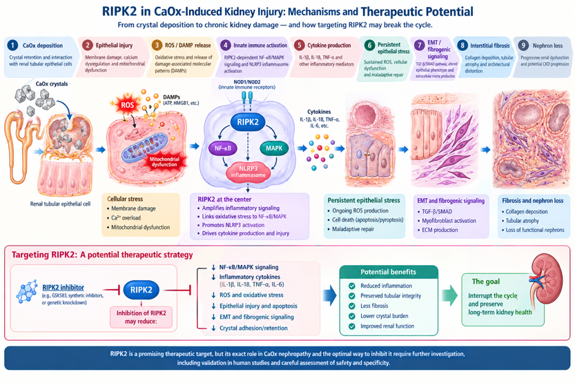
The notion that nephrocalcinosis merely represents a physical consequence of urine supersaturation or a precursor to nephrolithiasis has seen a drastic shift. The conventional view of calcium oxalate disease has largely focused on crystal formation, aggregation, adhesion, and urinary obstruction. However, latest evidence suggests that crystal nucleation, growth, aggregation, adhesion, and retention are influenced by urinary inhibitors, epithelial surfaces, tubular transport, and local tissue injury. Additionally, it has become evident now that crystals can directly interact with renal tubular epithelial cells and resident immune cells and that crystal uptake or surface interaction can disrupt cellular membranes, lysosomal integrity, intracellular calcium homeostasis, and mitochondrial function. These events promote the generation of reactive oxygen species (ROS) and release of damage-associated molecular patterns (DAMPs). Generation of ROS and DAMPs activate immune pathways; inflammatory cytokines then further compromise neighboring epithelial cells and potentially increase susceptibility to additional crystal adhesion and retention.Importantly, the capability of CaOx exposure to induceepithelial–mesenchymal transition (EMT) is particularly relevant. Evidence indicates that CaOx exposure can activate TGF-β-associated pathways and promote phenotypic changes consistent with EMT. Although the precise contribution of EMT to human renal fibrosis remains an area of continuing investigation, its association with chronic crystal-induced renal remodeling makes it an important mechanistic pathway to investigate. The role of CaOx crystals in triggering the fibrogenic cascade by upregulating markers such as vimentin, α-SMA, and collagen, ultimately resulting in excessive ECM deposition and tubulointerstitial fibrosis, suggests a pathologically significant damaging effect brought upon by the deposition of these crystals.
Our research findings linking CaOx crystal deposition in the kidney with oxidative stress, innate immune activation, mitochondrial dysfunction, and renal fibrosis through RIPK2 kinase signaling are particularly significant. These findings shift the therapeutic paradigm from merely preventing crystal formation toward protecting renal cells from the pathological consequences of crystal deposition. By targeting the molecular pathways that drive crystal-induced inflammation and tissue remodeling, this approach may open new avenues for preventing the progression of nephrocalcinosis and recurrent crystal-induced renal injury to chronic kidney disease.
In addition, our research group has identified Nesfatin-1 as a promising potential therapeutic agent that has demonstrated protective effects against CaOx-induced renal cellular injury. Its beneficial effects in experimental models suggest that Nesfatin-1 may help counteract crystal-induced cellular stress and preserve renal epithelial integrity. Together, these findings highlight two complementary therapeutic strategies: targeting RIPK2-mediated inflammatory signaling to interrupt the mechanisms underlying renal injury, and exploring Nesfatin-1-mediated cytoprotection to promote cellular resilience and recovery. Further mechanistic investigations and in vivo validation will be essential to establish their therapeutic potential and translational relevance in preventing long-term renal damage.
Despite major advances in understanding the underlying causes and consequences of calcium and other mineral depositions in the kidney, several important challenges remain. The central challenge is to move beyond preventing or removing crystals and develop therapeutic strategies that protect renal tissue and prevent chronic damage. Although current clinical interventions can remove stones and reduce their recurrence, they do not necessarily prevent ongoing renal epithelial injury or fibrosis, particularly when crystals accumulate intracellularly. Emerging evidence suggests that crystal-induced renal injury involves a complex network of interconnected signalling pathways. However, the interplay among reactive oxygen species (ROS), mitochondrial dysfunction, NF-κB and MAPK signalling, NLRP3 inflammasome activation, and pro-inflammatory cytokine release remains incompletely understood. Although inflammatory signalling represents a promising therapeutic target, its inhibition must be approached cautiously, as these pathways also contribute to normal host defence, tissue repair, and physiological homeostasis.
Another major challenge is to identify the factors that determine whether crystal deposition results in transient epithelial injury or progresses to persistent inflammation, impaired tissue repair, fibrosis, nephron loss, and chronic kidney disease (CKD). The observation that not all patients with nephrolithiasis or nephrocalcinosis develop progressive renal dysfunction suggests that additional determinants, including genetic susceptibility, metabolic differences, and variations in inflammatory and cytoprotective responses, may influence disease progression. Longitudinal studies and diverse, multicentre cohorts are therefore essential to identify high-risk populations and clarify the contributions of age, geographical variation, dietary factors, inherited disorders, and access to healthcare.
Our investigations of RIPK2 and Nesfatin-1 provide a potential framework for developing targeted therapeutic strategies. The key challenge now is to establish whether modulating RIPK2-associated inflammatory signalling and enhancing endogenous cytoprotective mechanisms can preserve renal structure and function despite crystal exposure. Elucidating the underlying causal mechanisms, demonstrating reproducible protection in relevant in vivo models, and validating these findings in human tissues and clinical cohorts will be crucial steps towards translating these approaches into effective disease-modifying therapies.
The field of nephrolithiasis and nephrocalcinosis research is transitioning from a predominantly physicochemical view of mineral deposition towards a more integrated understanding of crystal-induced cellular injury, inflammation, and tissue remodelling. Current clinical approaches are effective in removing stones and reducing recurrence in many patients, but strategies that directly prevent or reverse crystal-induced epithelial injury and renal fibrosis remain limited. Emerging evidence implicates oxidative stress, mitochondrial dysfunction, innate immune activation, and interconnected inflammatory signalling pathways as important contributors to disease progression. However, their causal relationships, relative contributions, and therapeutic relevance require further investigation. In this context, RIPK2-mediated inflammatory signalling and endogenous cytoprotective mechanisms involving Nesfatin-1 represent promising avenues for exploration, rather than established therapeutic solutions. The next major advance will depend on integrating mechanistic studies with robust preclinical validation, genetic and clinical investigations, and longitudinal assessment of renal outcomes. Ultimately, the field must move beyond understanding how crystals form towards determining how crystal-induced tissue damage can be prevented, limited, or reversed to preserve long-term kidney health.