Why VKH deserves a closer look
We chose to discuss Vogt–Koyanagi–Harada (VKH) disease because of its rarity, potential to cause severe visual loss, and the diagnostic challenges it may show during the initial presentation. In some cases of VKH, acute ocular pain accompanied by elevated intraocular pressure can mimic acute angle-closure glaucoma, potentially leading to an incorrect initial diagnosis. Prompt identification of VKH is crucial, as early initiation of appropriate systemic immunosuppressive therapy may help preserve visual function and limit complications involving the eyes, nervous system, auditory system, and skin.
VKH syndrome constitutes a multisystem autoimmune disorder, characterized by granulomatous inflammation targeting melanocyte-containing tissues, including the uveal tract, meninges, inner ear, and skin. Pathogenesis is attributed to T cell–mediated immune responses against melanocytic antigens in genetically susceptible individuals, with increased prevalence among patients of Asian, Hispanic, Middle Eastern, and Native American ancestry.
“VKH can mimic angle-closure glaucoma. Inflammation may push structures inside the eye forward and narrow the drainage angle.”
A disease that reaches beyond the eyes
Ocular: VKH primarily affects both eyes, producing granulomatous panuveitis and marked inflammation of the choroid. Accumulation of fluid beneath the retina may result in serous or exudative retinal detachments. Auditory: The inner ear may also be affected, particularly the melanocyte-containing structures. Patients can develop tinnitus, vertigo, and sensorineural hearing impairment. Neurological: In the early stage, inflammation of the meninges (coverings of the brain) may present with headache, fever, and neck stiffness. Cerebrospinal fluid examination may demonstrate an increased number of inflammatory cells. Cutaneous and hair changes: As the disease progresses, loss of pigmentation may become apparent. Typical findings include vitiligo, whitening of the eyelashes or eyebrows (poliosis), and areas of hair loss (alopecia). These changes are more characteristic of the later convalescent or chronic stages.
VKH generally progresses through four stages. The prodromal phase lasts for days to weeks and involves symptoms of malaise, headache, nausea, and/or neck and back stiffness, which was reported in about 50% of patients with VKH. The acute uveitic phase is characterized by photophobia, visual disturbance, ocular inflammation (granulomatous panuveitis), serous retinal detachments, and optic disc edema. In the convalescent phase, ocular inflammation gradually begins to subside while depigmentation of the fundus (“sunset-glow fundus”) becomes more evident. In the chronic or recurrent phase, persistent or recurrent episodes of granulomatous anterior uveitis and other ocular complications may occur.
A diagnosis revealed by looking deeper
The case published as “A rare case of Vogt-Koyanagi-Harada syndrome misdiagnosed as bilateral primary angle-closure suspect” is from a tertiary clinic in the eastern part of India. It describes a middle-aged Asian woman who initially received peripheral iridotomy for presumed primary angle-closure before posterior segment evaluation established it as VKH.
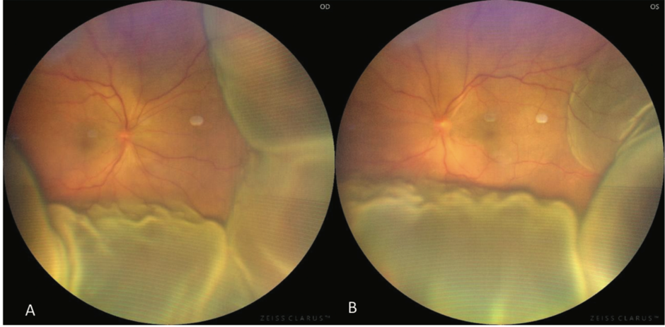
On further evaluation, she had bilateral shallow anterior chambers despite prior iridotomy, anterior chamber inflammation with pigmented keratic precipitates, optic disc edema, and multilobular serous retinal detachments. Posterior segment imaging showed focal areas of delayed choroidal perfusion with multiple pinpoint leaks giving an appearance of “starry sky” and late pooling. Optical coherence tomography (OCT) confirmed multiple serous detachments and a wavy retinal pigment epithelium. Systemic findings of cutaneous hypopigmentation and unilateral sensorineural hearing loss completed the clinical picture consistent with complete VKH.
High-dose intravenous methylprednisolone followed by oral steroids and azathioprine led to marked visual recovery and resolution of subretinal fluid. But the therapy was discontinued due to patient factors, which produced persistent subretinal fluid that resolved when immunosuppressive treatment was resumed. Subsequently, it led to the development of a “sunset-glow” fundus.
Lessons from a missed diagnosis
This case clinically instructs on several aspects. First, it highlights that VKH can clinically mimic primary angle-closure glaucoma (PACG) at presentation. Inflammatory choroidal thickening and ciliochoroidal effusion may shift the lens–iris diaphragm anteriorly, producing secondary angle-closure that is reversible with appropriate anti-inflammatory therapy. Failure to recognize posterior inflammatory signs risks unnecessary procedures such as iridotomy, as done in this case.
Second, the case reinforces that subtle anterior segment inflammatory signs (cells, keratic precipitates), bilateral symptoms, or posterior segment complaints should initiate posterior imaging before committing to any definitive angle-closure interventions.
Thirdly, from a therapeutic outlook, the favourable response from initiating high-dose systemic corticosteroids and the use of azathioprine for steroid-sparing maintenance are consistent with current practices: aggressive initial control of inflammation followed by sustained immunomodulation. This reduces relapse risk and chronic tissue damage. Lastly, the return of subretinal fluid after treatment discontinuation emphasizes the importance of patient counselling regarding the duration of therapy, adherence, and close monitoring. Follow-up to document visual function, choroidal thickness, and treatment toxicity is essential.
The clues that distinguish VKH from glaucoma
Bilateral involvement with systemic manifestations: VKH generally affects both eyes and may be associated with headache, tinnitus, hearing disturbances, or a recent flu-like illness. In contrast, PACG commonly begins in one eye and is not typically associated with systemic inflammatory symptoms. Posterior segment involvement: Findings such as diffuse thickening of the choroid, serous/exudative retinal detachment, and irregular alterations of the retinal pigment epithelium on OCT or ultrasonography are suggestive of VKH. In uncomplicated PACG, the posterior segment is not the primary site of disease. Cause of anterior chamber shallowing: A shallow anterior chamber can occur in VKH because inflammatory swelling of the choroid causes forward displacement of the uveal tissues. This differs from PACG, where angle closure is primarily related to anatomical crowding and pupillary block. Differences in treatment response: VKH is an inflammatory disorder and usually requires prompt systemic corticosteroid therapy and, when indicated, other immunosuppressive treatment. PACG, on the other hand, is managed by lowering intraocular pressure and opening the closed angle, often with medications and laser peripheral iridotomy.
Improving diagnosis and follow-up
To enhance educational value and reproducibility in future reports, it would be advantageous to incorporate anterior segment imaging such as ultrasound biomicroscopy (UBM) to objectively document ciliochoroidal effusion and the position of the lens–iris diaphragm. Additionally, providing a detailed account of the immunosuppressive regimen, including specific agents, dosing schedules, and monitoring protocols, would strengthen the clinical utility of the case. To summarise, this case serves as an important reminder that VKH should be included in the differential diagnosis of bilateral or atypical angle-closure scenarios. A thorough ocular and systemic evaluation, supported by multimodal imaging, timely initiation of systemic immunotherapy, and clear patient counselling regarding treatment adherence, are essential to preserving vision and minimizing the risk of chronic complications in VKH.