
Looking Beyond What We Can See
Plants are foundational to life on Earth, yet many aspects of their biology remain hidden. Over decades of research, we have revealed much about plant form and function, but understanding how plants survive changing and stressful environments still requires probing their processes at finer spatial and temporal scales. Imaging morphological and anatomical features with advanced microscopes has helped, but important limitations persist: rigid cell walls, densely light scattering tissues, and strong autofluorescence often obscure signals. Equally limiting is our chemical toolbox, we still lack many probes and sensors that can reliably detect the right molecules in the right place at the right time within living plant tissues. Progress in instrumentation has been steady, but the next major advances will likely come from chemistry. Next generation fluorescent probes, genetically encoded sensors, and small molecule reporters tailored for plant biology will let us track metabolites, ions, signaling molecules, and enzymatic activities with high spatial and temporal resolution. Coupling such probes with improved optical clearing methods, adaptive optics, and deeper penetration imaging modalities will overcome physical barriers and reveal dynamic processes in intact tissues. Together, better chemical tools and complementary imaging technologies will open new windows into plant development, stress responses, and adaptation mechanisms, and help us understand not just structure, but the molecular choreography that keeps plants alive.
The Biggest Unanswered Questions
Plant anatomy and biology have been advanced using fluorescent dyes, small-molecule probes, genetically encoded indicators, and self-labeling systems, although certain drawbacks leave unanswered questions and hinder further breakthroughs. As the field progresses, major challenges remain in visualizing the real-time dynamics of reactive oxygen species (ROS), ions, hormones, and metabolites; tracking the movement of signaling molecules; and monitoring cell wall and plasma membrane remodeling during development and stress responses. Further, physiological questions like tracing the assembly and dissolution of transient receptor ligand interactions, identification of in vivo ubiquitination, proteostasis, turnover mechanisms, apoplastic vs. cytoplasmic signaling dynamics, and hormonal crosstalk, among others, remain unanswered. A central challenge is bridging the gap between static structural and molecular predictions and the dynamic, real-time behavior of biomolecules in living plant cells. Achieving this goal will require next-generation probes that operate reliably in the chemically complex, highly autofluorescent environment of plant tissues while maintaining exceptional sensitivity, specificity, and spatiotemporal resolution.
Scientific and Technological Breakthroughs Needed
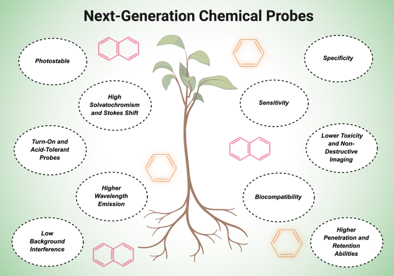
One of the major challenges in deciphering the structural and molecular complexity of plants lies in the design and synthesis of highly targeted probes and dyes. Although numerous fluorescent dyes and probes have been developed since the advent of modern plant biology, several limitations continue to hinder their broader application. These include insufficient cell- and tissue-specific selectivity, cytotoxicity, limited biocompatibility for long-term live-cell imaging, inadequate tissue penetration and retention, and suboptimal emission wavelengths. The development of probes with longer emission wavelengths, particularly near-infrared (NIR) fluorophores and two-photon probes, would minimize background interference and substantially enhance imaging performance in plant tissues.
Another major challenge is overcoming the inherent autofluorescence of plant tissues, which arises primarily from chlorophyll, lignin, and other cell wall-associated components that emit strongly within the visible spectrum. This issue could be addressed by developing NIR probes that operate in the infrared region or fluorophores with large Stokes shifts and pronounced solvatochromic properties, thereby enabling efficient spectral separation between endogenous autofluorescence and probe emission.
Another drawback is the widespread use of constitutively fluorescent probes, which emit continuously irrespective of biological activity, resulting in elevated background fluorescence until excess probe is removed. This limitation underscores the need for activatable, or “turn-on,” probes that fluoresce only in response to specific molecular interactions, biochemical reactions, or signaling events, thereby enabling higher imaging contrast and real-time visualization of dynamic cellular processes.
Next, the development of acid-tolerant probes for compartments such as the apoplast and vacuoles with low pH values remains an open avenue, given that dyes such as standard GFPs and pHluorins denature or quench their quantum yields at low pH. Also, GFP tags are too bulky to bind to small signaling peptides, phytohormones, lipids, and cell-wall glycans, underscoring the need for non-disruptive bio-orthogonal labeling via click chemistry, which enables their imaging without hindering natural binding affinities. A systematic transition from simple promoter-reporter constructs to genetically encoded biosensors and degron systems could pave the way for measuring real-time dynamic fluxes by quantifying fluorescence gain/loss. Further, measuring protein dynamics as an absolute measure of complete protein abundance can be advanced through the use of activity-based probes that chemically tag themselves at catalytic sites via targeted chemistries and trace specific localization patterns. Also, the use of genetically targetable chemical probes, as well as probes for phytohormones, enzymes, metabolites, and cell wall components, remains limited in terms of specificity. Expanding the repertoire of these probes, together with advances in fluorophore engineering, bioorthogonal chemistry, and biosensor design, will be essential for bridging the gap between static molecular observations and the dynamic, real-time visualization of plant physiology at cellular and subcellular resolution.
Emerging Trends Shaping the Future
Recent technological advances have transformed the way plant scientists investigate cellular chemistry, physiology, and tissue architecture. Among these developments, small-molecule fluorescent probes have enabled the sensitive detection of reactive oxygen species (ROS), ions, pH, and stress-associated biomarkers while facilitating live, non-destructive imaging. Likewise, genetically encoded indicators, self-labeling and hybrid-labeling strategies, and near-infrared (NIR) and two-photon fluorescent probes have emerged as powerful tools that address the increasing demand for high-resolution, real-time visualization of dynamic biological processes. Nevertheless, there remains a growing need for probes capable of monitoring cell wall composition, membrane chemistry and dynamics, and stress-responsive molecular events with greater specificity and spatiotemporal resolution. One particularly promising innovation is the development of plant tissue-clearing reagents such as ClearSee and ClearSeeAlpha, which efficiently reduce chlorophyll-mediated autofluorescence while preserving compatibility with a wide range of fluorescent dyes, proteins, and probes. These clearing agents overcome many of the limitations associated with conventional staining and clearing techniques, thereby improving imaging depth, signal quality, and tissue transparency.
Another significant advancement is the development of highly photostable fluorophores that minimize photobleaching, a long-standing limitation of conventional fluorescent dyes. An excellent example is MitoPB Yellow, a photostable fluorescent probe specifically designed to visualize the mitochondrial inner membrane. Its high selectivity, sensitivity, and resistance to photobleaching enable prolonged imaging with reduced background interference, making it particularly valuable for studying mitochondrial dynamics in living cells.
Similarly, the recently developed photostable fluorophore PhoxBright 430 (PB430) utilizes an intramolecular charge-transfer mechanism to achieve high solvatochromism, excellent quantum yields, and remarkable photostability. Its successful conjugation to antibodies has enabled stable fluorescence signals for long-term visualization of cytoskeletal dynamics, demonstrating the potential of this class of fluorophores for advanced live-cell imaging applications.
In concert with the development of these probes and stains, modern-day advanced microscopic techniques, such as but not limited to Super-Resolution microscopy, including SIM (Structured Illumination Microscopy) and STORM (Stochastic Optical Reconstruction Microscopy), Expansion microscopy (ExM), and Light Sheet Fluorescence microscopy (LSFM), have significantly provided a concerted enhancement in the imaging applications. These techniques have enhanced the optical resolution to the ~20-60 nm scale, reduced photobleaching, while maintaining high zoom and magnification capabilities, providing impetus for visualizing single-cell modules. These efforts on both the equipment and interface sides offer promising capabilities for scientific advancement.
How Will the Field Evolve?
The future of plant studies and innovations lies in the transition from general-purpose fluorescent staining molecules to highly tailored, synthetic molecular designs. The field seems to evolve along two paths: more specialized techniques that address the needs of specific probe compartmentalization, and towards specific signals and biological questions. In short, plant imaging must move from simply making things visible towards making the invisible biochemical dynamics measurable in living plants. This transformation will require probes capable of reporting real-time changes in molecular interactions, enzyme activities, signaling pathways, and metabolite dynamics rather than merely providing static localization information. Another avenue is to move from single static maps to multiplexed live biosensing, and from in vitro binding assays to in vivo quantitative approaches, including fluorescence resonance energy transfer (FRET), fluorescence lifetime imaging microscopy (FLIM), and other advanced fluorescence-based techniques coupled with next-generation chemical probes and genetically encoded biosensors. The move from the simple, general to the specific and complex would pave the way for an integral understanding of these systems and their development.
What Lies Ahead?
The major developments foreseen in this rapidly advancing field include the development of NIR and two-photon probes specialized for out-of-reach signaling molecules, including, but not limited to, phytohormones, ligand-receptor interactions, enzymes, and metal ions. The field would greatly benefit from organelle-targeted and protein-targeted dyes, as well as probes with biocompatibility, low toxicity, and background interference. In the coming years, the establishment of comprehensive probe libraries tailored for stress biology, cell wall dynamics, metabolite mapping, and cellular signaling will significantly expand our understanding of plant physiology and development. Such resources will facilitate the simultaneous investigation of multiple molecular processes, providing a more comprehensive view of plant responses to developmental and environmental cues. The major outlook for plant scientists would be a shift from traditional probes to more specific, targeted entities, requiring optimizations in well-studied plant systems, but would enhance the overall outlook and provide us with more information about their workings and dynamics.